Assoc. Prof. Naoki Suzuki, PhD. / Veterinarian (licensed in Japan), Dairy Ecosystem Research and Development Center
On February 1, 2013, Hiroshima University established two new programs: the “Distinguished Professors” (DP) program and the “Distinguished Researchers” (DR) program. Individuals who are part of these programs are recognized as senior and junior faculty members respectively, who are engaged in extraordinarily distinguished research activities.
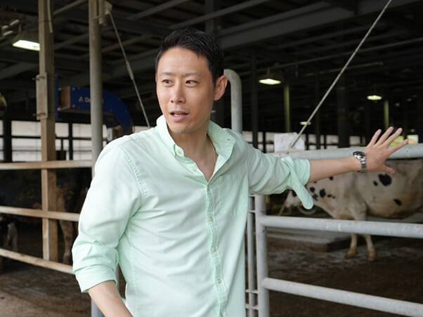
Rapid Infection Diagnosis for a Stable Supply and Safety of Livestock Products
Leveraging Vet Experience to Control Infectious Diseases in Livestock
My area of expertise is livestock hygiene, including things like infectious diseases. In particular, my research brings a bacteriological approach to on-farm control of infectious diseases caused by bacteria.
I was inspired to become a researcher as an undergraduate student when I was given an assignment that turned out to be challenging and very rewarding. It was part of a project led by the Ministry of Agriculture, Forestry and Fisheries, and I found it motivating to see my research outcomes being applied in real-life sanitation measures. Having said that, research has not been my one and only path since then. After graduating, I worked as a livestock veterinarian for seven and a half years, and that experience in clinical practice broadened my perspective and continues to inform my research work today.
A Rapid Diagnostic Technique for Mastitis in Dairy Cows
My research involves using basic bacteriology and applied microbiology to discover methods for on-farm control of infectious diseases, and my strength lies in harnessing clinical microbiology to achieve solutions that are highly relevant to farms. One example is the development of a rapid diagnostic technique for mastitis in dairy cows.
Mastitis is an inflammation of the mammary gland; it is found in around 20% of cows, making it a common headache for farmers. Mastitis also occurs among people, but in humans it is most commonly caused by clogging of the mammary gland, whereas bovine mastitis is defined as an inflammation of the mammary gland resulting from microbial infection.
Milk from udders with mastitis has to be discarded, resulting in tremendous economic losses. Therefore, when an abnormality is detected in milk or udders, prompt action is vital. The problem is that the microorganisms that cause mastitis are diverse. Not only are there many types of bacteria that can cause mastitis, such as Staphylococcus aureus and Escherichia coli, but fungi, algae, and other microorganisms can also cause the disease. Appropriate treatment and prevention requires identification of the microorganism, but conventional methods, which involve culturing and testing, take a whole day at the earliest, and sometimes up to a whole week. Also, because antimicrobials are used to treat infections caused by bacteria, the issue of antimicrobial resistance (AMR) is a constant concern. Antimicrobials are used in greater volumes in livestock than in humans, and the potential for inadvertently creating AMR in livestock poses a public health risk. This is why it is so important that antimicrobials be used appropriately and based on actual evidence.
Motivated by these challenges, I have been working since 2021 to develop a rapid diagnostic technique for mastitis and have come up with a technique that can diagnose the cause in less than a day. The technique uses gram staining to color-code bacteria suspended in milk into two groups—gram-positive and gram-negative—so they can be observed under a microscope. The gram staining method has not been widely used on bacteria in milk because the milk proteins and fats interfere with the image of the bacteria in the microscope when the milk is placed directly on a glass slide. By placing the milk in a centrifuge and separating out the bacteria, I am able to observe them at high sensitivity.
Gram staining itself cannot identify the type of bacteria, but the information it does yield is good enough because knowing if something is gram-positive or gram-negative makes it possible to choose a treatment plan and antimicrobials.
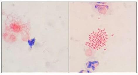
A micrograph of bacteria observed using gram staining showing gram-positive bacteria on the left (blue) and gram-negative bacteria on the right (red).
In addition, this technique has some unique features. Because milk contains large amounts of leukocytes derived from cows, and because the centrifugation separates these out together with the bacteria, this method makes it possible to observe both. You probably know that there are different types of white blood cells, such as neutrophils and lymphocytes—you’ve no doubt seen these listed in your health checkup results. The composition of these changes when you get an inflammation, which means that observing white blood cells provides a detailed picture of inflammation. In other words, this method of gram staining kills two birds with one stone, allowing bacteriological diagnosis and the degree of inflammation to be determined in the same microscopic image.
An App to Promote the Use of Diagnostic Technology
These days, I am eager to promote widespread uptake of this diagnostic technology. To that end, I travel throughout Japan and around the world showcasing the technology and providing hands-on training. At the same time, I am pursuing research to make this technology standard. Gram-stained bacterial imaging requires a knowledge of microbiology, but also an expert “eye.” To this end, I am working with an IT startup to develop an app that will harness AI to assist diagnosis by analyzing microscopic images of bacteria taken with a smartphone.
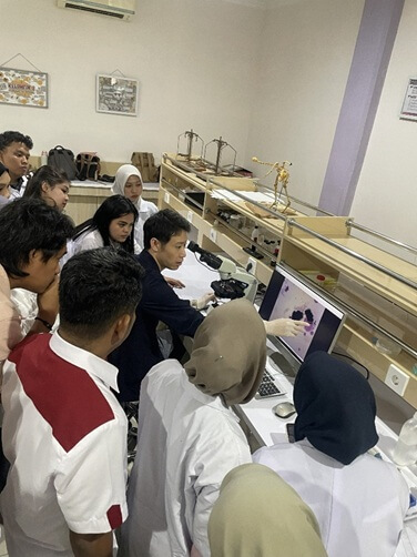
Practical training in Indonesia.
I hope to continue making a contribution to the stable supply and safety of food through development and basic research, such as simpler, farm-ready diagnostic technologies and automatic infectious disease detectors that can be incorporated into milking machines.
https://www.hiroshima-u.ac.jp/news/90206